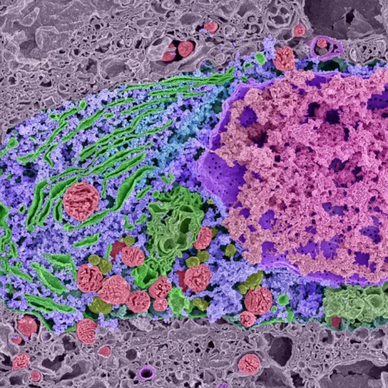
This is a cross section view of a single cell in the cortex of a rat’s brain. It was prepared using freeze fracture maceration, and imaged with scanning electron microscopy. Hundreds of tiled and depth stacked images were combined to create this mosaic.
The images were manually colourized to show the different organelles inside the cell. The DNA is in pink, nuclear envelope in purple, mitochondria in red, endoplasmic reticulum and golgi in green, and proteins in blue. Axons and dendrites around the cell are visible in violet.
This is the work I’m most proud of. Cells are incredibly beautiful beings, and this work shows the whole living structure, unifying the abstract visualizations of molecular biology into a coherent whole.