Zooming in on a diatom

2015
Microscopy

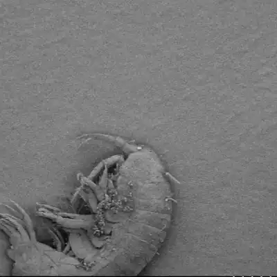
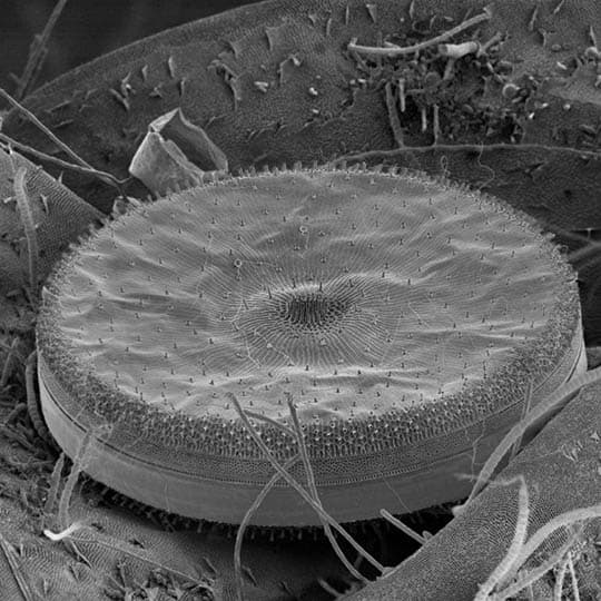
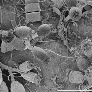
A bacterium on a diatom on an amphipod: that’s what is shown here on a This Diatom is named Bob, and it is the most photographed phytoplankton to ever live. This project aimed to capture the full beauty of Diatoms, microscopic algae with glass shells that live in all of […]

Gigapixel Diatom

2014
Microscopy

This Diatom is named Bob, and it is the most photographed phytoplankton to ever live. This project aimed to capture the full beauty of Diatoms, microscopic algae with glass shells that live in all of Earth’s aquatic environments. This species is Thalassiosira Rotula, a centric marine diatom that grows in […]

Microscopy Animations

2014
Microscopy

This video shows the microscopic world in a way seldom seen, with a 3D view. It shows various micro scenes of insects and marine life. These animations were produced with a Scanning Electron microscope, using a tedious manual process of imaging, tilting, readjusting, imaging again, and manual frame aligning. Each […]

The inside of a Neuron

2015
Microscopy

This is a cross section view of a single cell in the cortex of a rat’s brain. It was prepared using freeze fracture maceration, and imaged with scanning electron microscopy. Hundreds of tiled and depth stacked images were combined to create this mosaic. The images were manually colourized to show […]